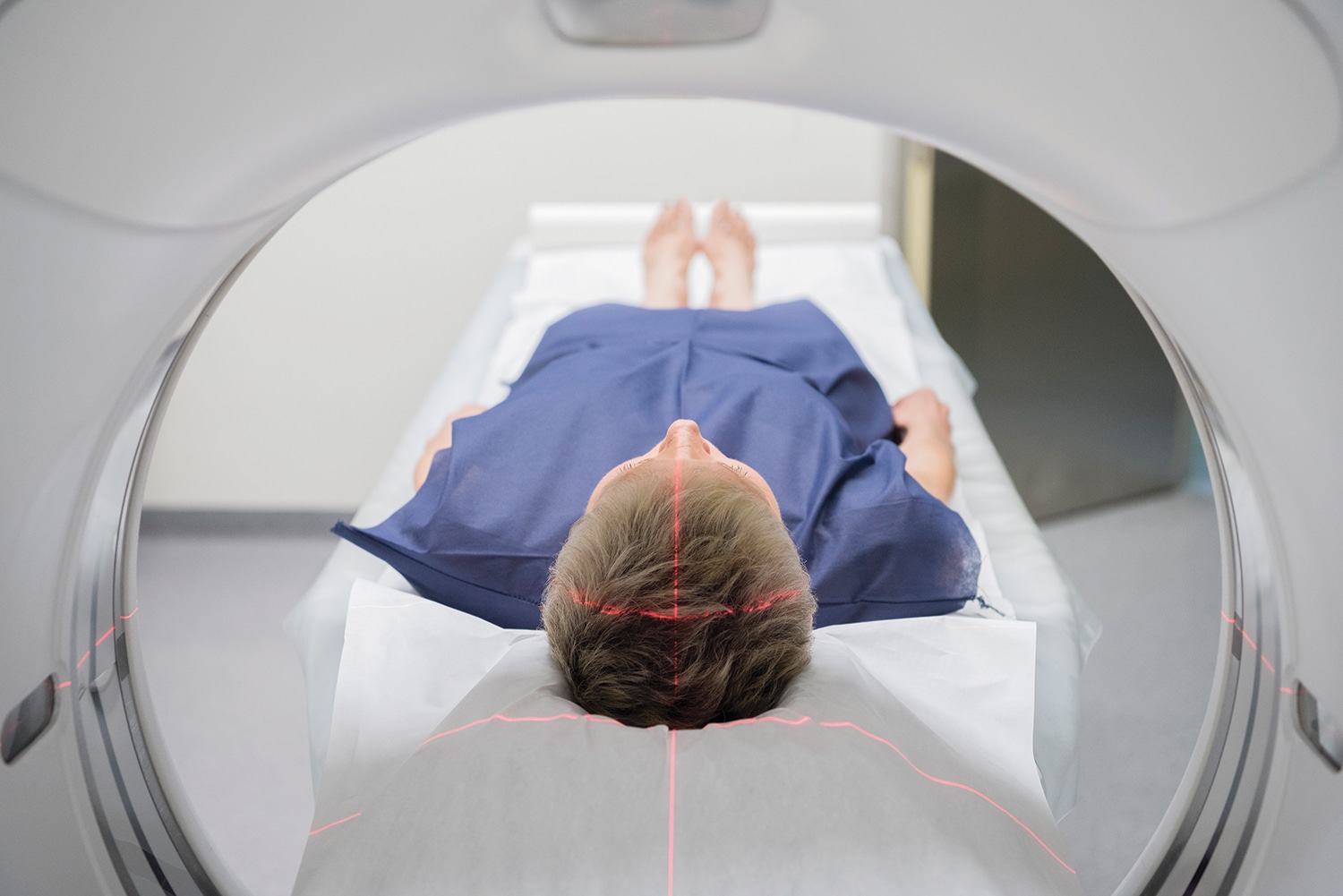
THE END OF CANCER TREATMENT can be a milestone for patients, but often it’s the beginning of a new phase of care: a post-treatment regimen of scans and other tests to look for signs of cancer recurrence. At intervals of three months, six months or longer, patients return for follow-up visits, often involving imaging scans, to find out if the cancer is back. Even if a scan shows no signs of cancer, patients know the clock will begin ticking toward the next round of tests.
Despite the importance of imaging in post-treatment testing, significant knowledge gaps exist among researchers and physicians, and discrepancies in clinical practice occur. More studies have scrutinized how to use imaging tests to detect cancer in the first place than have examined what tests to use and how often to look for recurrences once treatment is finished, according to cancer physicians and researchers who study imaging. And even when medical guidelines for post-treatment scanning are available, physicians don’t always follow them.
In a typical year, the average American will be exposed to 3 millisieverts (mSv) of radiation from sources such as radon in the home and cosmic radiation that penetrates the Earth’s atmosphere. A millisievert is a measure of the amount of radiation absorbed by the body. To explain the additional radiation in some imaging tests, the American Cancer Society compares them to typical annual exposure.
Chest X-ray: 0.1 mSv, or about 10 days of background exposure
Mammogram: 0.4 mSv, or about seven weeks
CT scan of the abdomen and pelvis: 10 mSv, or more than three years
PET/CT scan: 25 mSv, or about eight years
One concern with post-treatment imaging among doctors and patients is radiation exposure, a cancer risk factor. Doses for imaging tests generally vary from quite low for mammograms and chest X-rays to far higher for PET/CT scans. (MRI and ultrasound scans don’t use radiation.) One 2009 study, which looked at CT scan use for any reason, projected that if those rates of scanning continued, the result could be 29,000 more future cancers annually.
“For years there has been a pervasive attitude that ‘well, the patient has had cancer already. So, what’s the concern of giving them a little bit more diagnostic radiation?’” says Benjamin Franc, a radiologist at the Stanford University School of Medicine in California. In some scenarios—for example, when patients have been treated for aggressive malignancies—that is a “very valid point,” says Franc, who recently published a study showing wide geographic variation in post-treatment imaging of breast cancer patients. The benefits of potentially catching a recurrence earlier, even if more frequent imaging is required, likely outweigh concerns about lifetime radiation exposure, he says. In addition, older individuals are not considered to be as vulnerable to radiation exposure given that radiation-linked malignancies typically require decades to develop after exposure.
There are other costs for patients to shoulder besides radiation exposure, however. Higher deductibles are becoming more common, which means that even insured patients might need to foot a larger chunk of the imaging bill. Then there is “scanxiety,” the days and weeks of worry leading up to each scan. And sometimes scans capture spots or shadows that generate new questions and create the need for further decisions: Is more imaging or a biopsy needed to figure out what is going on, or should the individual simply wait to see what else develops?
“I think that there’s a perception that medical imaging is a panacea and that it only can be a benefit, when in fact there can be potential benefits and potential harms,” says Rebecca Smith-Bindman, a University of California, San Francisco radiologist who has studied radiation exposure and medical imaging. “And [the harms] are not trivial.”
For Julie Wrigley, a routine CT scan in 2016 shook up her life. Wrigley was roughly two years past the end of her treatment for stage IIIB colon cancer when a surveillance CT scan—quickly followed by a bone scan recommended by her doctor—revealed several potentially worrisome spots. “I ended up quitting my job completely,” says the mother of three from Anchorage, Alaska, who was then 42 years old and an attorney in a small firm. “I figured that if there was something in fact going on in my body that had gone to my bones, I wanted not to be working.”
Gaps in Research and Practice
Several factors have stymied attempts to research best practices for post-treatment imaging, says Stephanie Lee, a hematologist at Seattle’s Fred Hutchinson Cancer Research Center who studies cancer survivorship. Patients diagnosed with the same malignancy might differ in the aggressiveness of their cancer and in the treatment they receive, among other factors, making it difficult to recruit a uniform group to study. Also, patients in clinical trials often must agree to be randomized to different imaging approaches—a tough sell given that some patients might end up getting less imaging than they would prefer for their peace of mind, Lee says.
Evidence from studies on post-treatment imaging is lacking, so what are recommendations based on? Smith-Bindman says that sometimes they are drawn from expert consensus by cancer physicians. In other cases, guidelines are based on insights gained from research studies in which patients receive scans regularly after the end of treatment to track the ongoing effectiveness of drugs and other therapies. Another tactic is to take findings from studies looking at the best way to screen for cancer and apply them to post-treatment recommendations, says David Gerber, an oncologist and lung cancer specialist at the University of Texas Southwestern Medical Center in Dallas.
For instance, guidelines previously said that either a chest X-ray or a low-dose CT scan could be used to monitor for lung cancer recurrence. Then the National Lung Screening Trial determined that CT scans were better at screening for malignancy, and guidelines for post-treatment surveillance subsequently followed suit, Gerber says. “But there’s never been a large study to prove that’s the case,” he says of the conclusion that CT scans are better than X-rays to monitor for recurrence. “That’s us extrapolating from screening trials.”
Even when guidelines include specific post-treatment imaging recommendations, Smith-Bindman says, physicians don’t always follow them. Franc’s study, in the July 2018 Journal of the National Comprehensive Cancer Network, illustrated geographic gaps between guidelines and actual surveillance for 36,045 women with low-risk breast cancer. The American Society of Clinical Oncology (ASCO) and the National Comprehensive Cancer Network recommend mammograms to look for cancer after treatment in women without symptoms who have a low risk for recurrence. ASCO guidelines state that CT and PET scans should be avoided in this low-risk group of breast cancer survivors. The rationale: Studies haven’t shown any benefit, and the imaging can lead to false-positive results that can drive additional procedures and anxiety.
Yet nearly one-third of the women in the study had at least one high-cost imaging procedure, such as a CT or a PET scan, in the first 18 months after their breast cancer surgery. A CT scan can be used alone or in combination with a PET scan.
One key question is how often imaging should be done, says George J. Chang, chief of colon and rectal surgery at the University of Texas MD Anderson Cancer Center in Houston. Chang led a study involving more than 8,500 colorectal cancer patients that tracked how long it took to detect a cancer recurrence between two groups of patients, one of which got an average of 2.9 imaging scans along with blood testing during the first three years of surveillance and the other which averaged 1.6 imaging scans plus bloodwork. In the high-intensity surveillance group, it took a median of 15.1 months to detect a recurrence compared with 16 months in the low-intensity group, according to findings published in the May 22/29, 2018, JAMA.
“A lot of times patients are thinking ‘if I get more frequent testing, I’m going to pick it up sooner,’” Chang says. “But our study would show that that actually didn’t bear out.” Most importantly, he says, there was no difference in overall survival between the two patient groups.
Ask some questions prior to receiving the next imaging test.
Radiologist Rebecca Smith-Bindman of the University of California, San Francisco, advises patients to discuss post-treatment imaging recommendations with their physicians, particularly if they are concerned about radiation exposure. “I wouldn’t assume, which is really hard for a lot of patients, that more imaging is better,” she says.
Among the questions she suggests patients ask their doctors are:
- What do you expect to learn from the test? The goal of imaging is to find a recurrence before symptoms develop and while the malignancy is still treatable, Smith-Bindman says.
- Are there research data or guidelines supporting how often you want me to undergo this imaging test?
- Which type of test will have the fewest side effects, including those related to radiation exposure?
- Can I replace this imaging test with an ultrasound? For surveillance after some cancers, such as ovarian cancer, this might be an option, Smith-Bindman says.
- Is there a way to conduct this test with a low radiation dose? For instance, Smith-Bindman says, a CT scan of some organs such as the liver can be completed with a single scan or multiple scans in a single session, with the multiple-scan approach boosting radiation exposure.
Radiation and Other Anxieties
When weighing the pros and cons of an imaging test, patients should keep in mind not only that each type of imaging carries different radiation exposure, but also that there can be variations in exposure depending upon the facility and how a scan is administered, Smith-Bindman says. She was involved with research published in 2009 that looked at CT radiation doses for parts of the body including the head, neck and abdomen at four institutions and found that the largest radiation dose was on average 13 times greater than the smallest dose for each type of CT scan.
Moreover, some CT scans capture an area far bigger than the organ that is the focus of the scan, says Gerber. For instance, he says, a chest CT scan to check for a lung cancer recurrence may also include the stomach, kidneys, esophagus, heart, breasts, thyroid gland and at least part of the liver. Thus, the scan might reveal potential abnormalities in other areas that may or may not be life-threatening, Gerber says. Once something worrisome is identified, it has to be monitored to see if it changes, or the area has to be biopsied, which carries surgical risks, he adds.
For Wrigley, the colon cancer survivor living in Alaska, that seemingly routine CT scan led to the bone scan and the unsettling identification of several troubling spots: one partway down her spine, another on her shoulder bone and a third on her temple. She consulted with many doctors, including her own husband, who is a physician, and worried that the next step would be a bone biopsy, which she jokes “would involve sticking needles into places I didn’t want to.” Instead, she decided to wait a year and get scanned again to see whether the spots had changed. After quitting her job to spend more time with her family, Wrigley turned to exercise and meditation. Did she ever think about those spots?
“All the time,” she says immediately. “All the time it was in the back of my head. You can’t really get rid of it.” That is, until another CT scan the following year showed no signs of change.
Nearly three years after she first found out about the spots, Wrigley continues to worry about long-term radiation exposure as her scans accumulate. But she admits that she’ll sometimes seek out an additional CT scan that’s not part of her regular imaging schedule if gastrointestinal pain or distress—a side effect of surgical removal of her tumor and part of her colon—is persistent. “You think that if you just get that scan, then you’ll know if something is going on or not, and you can sleep,” she says.
But even though a “clean” scan brings relief, it’s difficult not to wonder again as the months pass whether cancerous cells are developing, Wrigley says. “I think I’ll always try to be aware of what my body is saying,” she says. “And I am certain it will lead to more scans than I would prefer.”
Cancer Today magazine is free to cancer patients, survivors and caregivers who live in the U.S. Subscribe here to receive four issues per year.


